Treatment Planning
Another significant benefit of cone beam X-rays is their ability to assist in treatment planning. With a detailed three-dimensional image, dentists can better plan for complex procedures such as dental implant placement, orthodontic treatment, and oral surgery. By studying the patient's dental structures from all angles, dentists can map out the most effective treatment approach, minimizing potential complications and ensuring successful outcomes.
Improved Patient Communication
Cone beam X-rays not only benefit dentists, but they also improve patient communication. With a clear and highly visual representation of their oral health, patients can better understand their conditions and the recommended treatment options. Dentists can use the cone beam X-ray images to educate patients on the need for specific treatments, enhancing their understanding and cooperation. This not only promotes better patient satisfaction but also facilitates shared decision-making, leading to more successful treatment outcomes.
Reduced Radiation Exposure
Despite providing more comprehensive images, cone beam X-rays actually expose patients to lower radiation levels compared to traditional computed tomography (CT) scans. This is because cone beam X-rays use a focused X-ray beam closely aligned with the area of interest, resulting in more precise imaging with less scatter radiation. Dentists can therefore obtain detailed information about dental structures while minimizing radiation exposure, making cone beam X-rays a safer alternative.
Increased Efficiency
Lastly, cone beam X-rays offer exceptional efficiency in dental practices. With their fast scanning capabilities, dentists can obtain high-quality images in a matter of seconds. This saves valuable time for both the patient and the dental team. Additionally, cone beam X-rays eliminate the need for traditional film development, reducing the wait time for results. Immediate access to detailed images improves overall workflow and allows dentists to make more informed decisions promptly.
In conclusion, cone beam X-rays have transformed the field of dentistry by providing accurate diagnoses, aiding in treatment planning, improving patient communication, reducing radiation exposure, and increasing efficiency. As this technology becomes more widely available, it will continue to enhance dental care by enabling dentists to deliver personalized treatments with improved precision and safety.
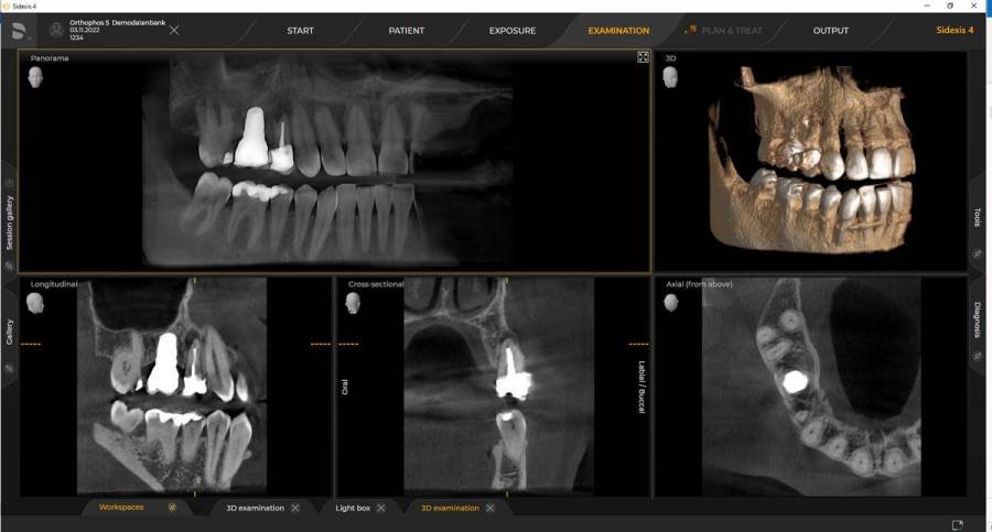
At the Dentist Group we utilize the Sirona Orthopus.